Laser Microscopy

Our range of services

To develop new components, the semiconductor industry has to accurately characterize them both structurally and electronically. Conventional optical analysis techniques are principally suitable for investigating these properties, but their diffraction-limited spatial resolution is not sufficient for modern semiconductor structures. By contrast, laser microscopy, as scattering near-field optical microscopy (SNOM) with broadband lasers, is very well suited for corresponding analyzes. Fraunhofer ILT has developed a broadband tunable mid-infrared laser system that opens up new spectral ranges and can be used to investigate stresses in industrial gallium nitride, for example. Likewise, it can investigate doping concentrations or free charge carriers in different materials.

Nanocomposite materials can be studied with laser microscopy as can commercial consumer products, such as nanoparticle-added cosmetics. In addition to near field microscopy, the institute uses other technologies, including confocal laser scanning microscopy, multiphoton microscopy, fluorescence lifetime imaging (FLIM) and polarization microscopy, as well as combinations of fluorescence and Raman microscopy.

The range of services Fraunhofer ILT offers includes feasibility studies, application-specific investigations with various microscopy methods, the development of laser-based processes, for example for material analysis, as well as individual consultation.

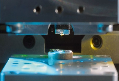
Near-field microscopes are suitable for investigations of modern semiconductor structures.
© Fraunhofer ILT, Aachen, Germany.
Near-field microscopes are suitable for investigations of modern semiconductor structures.
Near-field microscope, detailed view.
© Fraunhofer ILT, Aachen, Germany.
Near-field microscope, detailed view.
Live cell imaging of tissue cultures.
© Fraunhofer ILT, Aachen, Germany.
Live cell imaging of tissue cultures.

Laser Microscopy

  • Confocal laser scanning microscopy (CLSM)
  • Multiphoton microscopy
  • Fluorescence lifetime imaging (FLIM)
  • Polarization microscopy
  • Combination of fluorescence and Raman microscopy
  • Microscopic analysis of chemical reactions
  • Scanning near-field optical microscopy (SNOM) with broadband lasers

Branches

Laser technology can solve demanding tasks in many different industries. Whether as a tool in automotive production, as measuring equipment in the environmental sector, as a diagnostic or therapeutic instrument in medical technology or as a communication medium in space technology, the laser provides multiple uses with high productivity and high efficiency.

Read up about the innovations of the Fraunhofer ILT in a few selected industries and convince yourself!


Research with us!

Please do not hesitate to contact us if you have any questions about general topics! Our contact persons are happy to get in touch with you.

Publications

Bensmann, S., Gaußmann, F., Lewin, M., Wüppen, J., Nyga, S., Janzen, C., Jungbluth, B., Taubner, T.:
Near-field imaging and spectroscopy of locally strained GaN using an IR broadband laser
Opt. Expr. 22 (19), 22369-22381 (2014)

Bozkurt, A., Lassner, F., O'Dey, D., Deumens, R., Böcker, A., Schwendt, T., Janzen, C., Suschek, C., Tolba, R., Kobayashi, E., Sellhaus, B., Tholl, S., Eummelen, L., Schügner, F., Damink, L., Weis, J., Brook, G., Pallua, N.:
The role of microstructured and interconnected pore channels in a collagen-based nerve guide on axonal regeneration in peripheral nerves
Biomaterials 33, 5, S.
2012

Kaul, R. A., Mahlmann, D. M., Loosen, P.
Mach-Zehnder interference microscopy optically records electrically stimulated cellular activity in unstained nerve cells
J. Microsc. 240, Pt 1, 60-74
2010

Schwendt, T., Michalik, C., Zavrel, M., Dennig, A., Spiess, A. C., Poprawe, R., Janzen, C.
Determination of temporal and spatial concentration gradients in hydrogel beads using multiphoton microscopy techniques
Appl. Spectrosc. 64, Nr 7, 720-726
2010

A. C. Spiess, M. Zavrel, M. B. Ansorge-Schumacher, C. Janzen, C.Michalik, T. W. Schmidt, T. Schwendt, J. Büchs, R. Poprawe, W. Marquardt
Model discrimination for the propionic acid diffusion into hydrogel beads using lifetime confocal laser scanning microscopy
Chem. Eng. Sci.
63, 3457-3465, 2008

C. Janzen, A. Lenenbach, P. Jander, R. Noll
Neue Lasermesssysteme für bioanalytische und klinisch-diagnostische Aufgabenstellungen
VDI-BERICHTE
2011, 307-314, 2008

D. M. Mahlmann, J. Jahnke, P. Loosen
Rapid determination of the dry weight of single, living cyanobacterial cells using the Mach-Zehnder double beam interference microscope
Eur. J. Phycol.
43 Nr 4, 355-364, 2008

Our services cover a wide range of topics. Related topics to laser microscopy and further research and development focuses can be found under the following links.